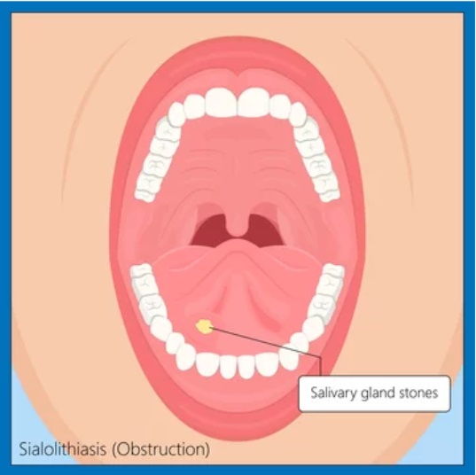
Salivary stones, medically known as SIALOLITHIASIS, can be a very painful and bothersome condition, affecting the salivary glands and hindering the natural flow of saliva.
What are Salivary Stones?
There are main 3 pairs of salivary glands in the human body —
The exact reason why these stones are formed is uncertain, however –
- PAROTID GLANDS ( The name of the parotid gland duct is Stensen Duct)
- SUBMANDIBULAR GLANDS ( The name of the Submandibular gland duct is Wharton’s Duct)
- SUBLINGUAL GLANDS
- Salivary secretions stasis,
- Inflammation of the salivary gland duct and
- Salivary duct injury is a significant contributing factor.
80-90% of calculi develop in Wharton’s duct of the submandibular salivary gland.
Stensen’s duct of the parotid gland constitutes 10–20%, and the sublingual duct only 1%.
The reasons why calculi mainly develop in Wharton’s duct of the submandibular salivary gland are the following –
- Wharton’s duct is longer and has a larger caliber.
- Wharton’s duct is angulated against gravity as it courses around the mylohyoid muscle.
- Submandibular secretions are more viscous
- Submandibular secretions have higher calcium and phosphorus concentration.
- Parotid stones are primarily located at the hilum or parenchyma, while in the submandibular gland, they tend to develop in the duct.
Composition of Salivary Stones –
They are composed mainly of calcium phosphate and carbonate combined with an organic matrix of glycol proteins, mucopolysaccharides, and small amounts of other salts such as magnesium, potassium, and ammonium.
Clinical features of SALIVARY STONES –
- Recurrent episodes of postprandial salivary colic pain and swelling.
- Past history of recurrent attacks of acute suppurative sialadenitis might be present.
- Bimanual palpation reveals the presence of a stone in most cases.
- Parotid stones might be seen just at the orifice of Stensen’s duct or along its course.
WHAT ARE THE TREATMENT OPTIONS?
Nonsurgical management of Salivary Stones consists of the use of –
- Sialogogues (are foods that increase salivary production)
- Local heat
- Hydration
- Massaging of the involved gland
- Antibiotic coverage is started in cases of infection.
- Manually milking out: Submandibular stones nearer the duct orifice may be manually milked out through the duct opening
Surgical management of Salivary Stones consists of –
1) Incision of duct:
Submandibular stones, which are no more than 2 cm from the duct orifice, may be either manually milked out through the duct opening or the duct is incised directly over the stone. There is no need for closure of Wharton’s duct after the procedure.
2) Sialadenectomy:
Submandibular stones located more proximal and near the gland will require sialadenectomy, which means surgical removal of the whole of the submandibular gland.
Parotid stones are more difficult to manage because of the anatomy of Stensen’s duct.
3) Recent advances:
Use of various combinations of baskets, graspers, and intracorporeal lithotripsy have been employed to treat sialolithiasis in both the parotid and submandibular glands.
4) Extracorporeal shock wave lithotripsy
It reduces stones to tiny fragments, which are then flushed out of the duct with spontaneous salivation or a secretagogue.
5) Sialoendoscopy:
Flexible endoscopes are used to visualize and remove salivary duct stones.
The Rise of Sialendoscopy: A Minimally Invasive Marvel
1. Introduction to Sialendoscopy
Sialendoscopy, a minimally invasive procedure, has emerged as a breakthrough in treating salivary stones. Unlike traditional methods, sialendoscopy allows for direct visualization and removal of stones within the salivary ducts, ensuring a more precise and effective intervention.
2. How Sialendoscopy Works
During sialendoscopy, a thin, flexible tube equipped with a camera is inserted into the salivary duct, providing a real-time view of the affected area. This allows the medical professional to precisely identify and remove the stone, minimizing damage to surrounding tissues. The procedure is performed on an outpatient basis, reducing the need for hospitalization and ensuring a quicker recovery.
3. Benefits of Sialendoscopy
- Sialendoscopy offers numerous advantages over traditional treatments.
- It is less invasive, resulting in reduced pain and swelling post-procedure.
- Additionally, the risk of complications is significantly lower compared to open surgery.
- Patients can expect a quicker recovery, enabling them to resume their daily activities promptly.
The Competitive Edge: Why Sialendoscopy Outperforms Traditional Methods
1. Precision and Accuracy
The key differentiator of sialendoscopy lies in its precision and accuracy.
By directly visualizing the stone, healthcare professionals can target and remove it with minimal disruption to surrounding tissues.
This ensures a more successful outcome than traditional methods, where the stone’s exact location may be challenging to ascertain.
2. Minimized Risks and Complications
Traditional surgical interventions for salivary stones come with inherent risks, including infection, bleeding, and damage to adjacent structures.
Sialendoscopy, being a minimally invasive procedure, significantly reduces these risks, making it a safer and more attractive option for patients seeking long-term relief from salivary stones.
3. Faster Recovery Times
With sialendoscopy, patients experience shorter recovery times compared to traditional surgery. The procedure’s minimally invasive nature allows for quicker healing, enabling individuals to return to their normal activities sooner.
Ensuring Success: The Role of Expertise in Sialendoscopy
1. Choosing a Skilled Sialendoscopist
While the benefits of sialendoscopy are evident, the procedure’s success depends on the expertise of the medical professional performing it.
Patients are encouraged to seek out experienced sialendoscopists with a proven track record in salivary stone management.
2. Patient Candidacy for Sialendoscopy
Not every individual with salivary stones may be an ideal candidate for sialendoscopy.
Factors such as stone size, location, and the overall health of the patient are crucial considerations.
A thorough evaluation by a qualified ENT specialist will determine the suitability of sialendoscopy for each case.
Conclusion: Embracing a Future Without Salivary Stone Worries
In conclusion, sialendoscopy stands as a beacon of hope for individuals grappling with the discomfort of salivary stones. Its precision, minimally invasive nature, and faster recovery times make it a superior choice over traditional treatment methods. As the medical community continues to refine and advance sialendoscopy techniques, the outlook for those suffering from salivary stones is increasingly optimistic. Embrace a future without the worries of salivary stones – explore the transformative benefits of sialendoscopy today.
THANK YOU
MEDICAL ADVICE DISCLAIMER:
This blog, including information, content, references, and opinions, is for informational purposes only.
The Author does not provide any medical advice on this platform.
Viewing, accessing, or reading this blog does not establish any doctor-patient relationship.
The information in this blog does not replace the services and opinions of a qualified medical professional who examines you and prescribes medicines.
If you have any medical questions, please refer to your doctor or qualified medical personnel for evaluation and management at a clinic/hospital near you.
The content provided in this blog represents the Author’s interpretation of research articles.
